CD138 + Cyclin D1
$795.00
Description
Product Description
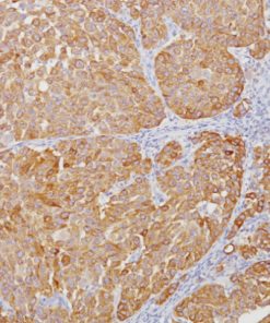
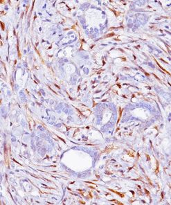
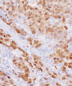
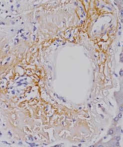
Detection of abnormal numbers and/or distribution of bone marrow (BM) plasma cells (PCs) on trephine biopsies can be of importance in the differential diagnosis of multiple myeloma (MM) and other PC neoplasms (1,2). CD138 expression in hematologic malignancy in bone marrow biopsy specimens is limited to nonneoplastic and neoplastic plasma cells and lymphoplasmacytic lymphoma in which strong membrane staining of neoplastic plasma cells is a consistent finding. Other hematopoietic malignancies are CD138 negative (1,2). Some marrow elements, such as stroma, mast cells, histiocytes, fat cells, osteoblasts and osteoclasts, and cells of hematopoietic lineage, are uniformly negative (1,2). Essentially all plasma cells are delineated with a strong membranous immunoreactivity, providing a highly sensitive and specific marker for quantitation of plasma cells in bone marrow biopsies. The reactivity for CD138 is well preserved in both Zenker-fixed and formalin-fixed decalcified tissue biopsies and the use of antigen retrieval do not alter the reactivity of CD138 (1,2). These results demonstrate that CD138 is a highly sensitive and specific marker that is useful for the rapid and precise localization of nonneoplastic and neoplastic PCs on routine BM sections. In addition, because of its clear-cut cell membrane localization, CD138 can be used in double-marker immunostaining reactions to evaluate precisely diagnostic nuclear markers such as Ki67, Cyclin D1, and Cyclin D3 in MMs. Immunohistochemistry (IHC) and fluorescence in situ hybridization (FISH) are used to investigate Cyclin D1 expression and the presence of chromosome 11 abnormalities in 48 MM patients (40 at diagnosis and 8 at relapse) and in normal plasma cells and hematopoietic precursors in BM biopsies (4). Cyclin D1 overexpression occurred in 12/48 (25%) of cases; combined IHC and FISH analyses in 39 patients showed Cyclin D1 positivity in all of the cases (7/7) bearing the t(11;14), in two of the 13 cases with trisomy 11. Normal plasma cells and other hematopoietic components in BM do not react to Cyclin D1 antibody. The data indicate that the t(11;14) translocation in MM leads to Cyclin D1 overexpression and that IHC analysis may represent a reliable means of identifying MM patients with the t(11;14) and, in general, of demonstrating the involvement of this gene in MM (4). The co-expression and visualization of CD138 (membranous) and Cyclin D1 (nuclear) in neoplastic plasma cells showing by double IHC staining make quantification of the tumor cells much easier to observe compared to individual slide interpretation (2,4).
Datasheets & SDS
| Download DS Data Sheet |
| Download SDS Sheet |
Browse more documents for this product (IFUs, datasheets, translations, SDS, and more).
References
1. Chilosi M, et al. CD138/syndecan-1: a useful immunohistochemical marker of normal and neoplastic plasma cells on routine trephine bone marrow biopsies. Mod Pathol. 1999;12:1101-6.
2. Yokoi S, et al. Cytogenetic study and analysis of protein expression in plasma cell myeloma with t(11;14)(q13;q32): Absence of BCL6 and SOX11, and infrequent expression of CD20 and PAX5. J Clin Exp Hematop. 2015;55:137-43.
3. O’Connell FP, et al. CD138 (Syndecan-1), a plasma cell marker immunohistochemical profile in hematopoietic and nonhematopoietic neoplasms. Am J Clin Pathol. 2004;121:254-63.
4. Pruneri G, et al. Immunohistochemical analysis of cyclin D1 shows deregulated expression in multiple myeloma with the t(11;14). Am J Pathol 2000, 156:1505-13.
5. Center for Disease Control Manual. Guide: Safety Management, NO. CDC-22, Atlanta, GA. April 30, 1976 “Decontamination of Laboratory Sink Drains to Remove Azide Salts.”
6. Clinical and Laboratory Standards Institute (CLSI). Protection of Laboratory Workers from Occupationally Acquired Infections; Approved Guideline-Fourth Edition CLSI document M29-A4 Wayne, PA 2014.
Related products
Primary Antibodies


