Meeting the Mark: Desmoglein 3 + p40 + Napsin A
Lung cancer is the leading cause of cancer-related deaths in the United States and worldwide, with non-small cell lung cancer (NSCLC)
accounting for 80% of lung cancers.1 NSCLC’s predominate subtypes are adenocarcinomas (ADC) and squamous cell carcinomas (SCC).1
Therapy of NSCLC depends on accurate histological classifications of these two subclassifications.1 Differentiating the two can be difficult,
especially when most lung tissue samples are collected through fine needle aspiration biopsies.1 Small biopsies such as this may only provide
a few tumorous cells or provide poorly differentiated cell morphologies.1 Immunohistochemistry (IHC) examination may provide clarification
and assist in classifying NSCLC.
Biocare’s Desmoglein 3 + p40 + Napsin A antibody cocktail is a composed of two mouse monoclonal antibodies and a rabbit monoclonal
antibody intended for use in the qualitative identification of Desmoglein 3, p40 and Napsin A proteins by IHC. Desmoglein 3 (DSG3) is a
calcium-binding transmembrane glycoprotein component of desmosomes in vertebrate epithelial cells.2,3 Desmoglein exhibits membranous
expression and connects with cytokeratins through desmoplakins and plakoglobin. DSG3 is particularly important in the cellular adhesion of
squamous epithelium, and as a result, it is often highly expressed in various squamous cell carcinomas (SqCC).4 Another highly specific SqCC
marker is p40. Biocare’s p40 clone [BC28] is a nuclear marker that recognizes an epitope unique to p40, which may result in diminished
reactivity in lung ADC and increased specificity.5-8 This combination of membrane and nuclear staining of DSG3 and p40, respectively, may
increase overall sensitivity for lung SCC and, in some cases, aid the pathologist with difficult specimens.5,9 The third component, Napsin A, is
a pepsin-like aspartic proteinase that is expressed in most lung ADCs 4-6 Studies have shown that Napsin A is a more sensitive and specific
marker than TTF-1 and is extremely specific for lung ADC versus lung SCC.4-6 Using innovative multiplex detection systems, such as Biocare’s
Mach 2 Double Stain 2 polymer, this cocktail of antibodies can be targeted and labeled simultaneously with two separate enzymes. With this
polymer, the membranous (DGS3) and nuclear (p40) mouse antibodies are labeled with an HRP enzyme and visualized with DAB, while the
rabbit antibody (Napsin A) is labeled with an AP enzyme and visualized with a Fast Red chromogen.
Lung carcinomas, particularly NSCLCs, are a mixed group of tumors exhibiting differences at the histologic, immunophenotypic, and molecular
genetic levels.1 The accurate classification of NSCLC is becoming increasingly important due to advancements in therapies.1Research has
shown triple marker cocktails have comparable sensitivities and specificities to individual markers.1 Ao et al. found Biocare’s cocktails to be
cost-efficient and may aid in conserving valuable tumor tissues during the immunohistochemical study and the subclassification of NSCLCs.1
Looking to maximize the diagnostic ability of a single needle biopsy? Biocare’s Desmoglein 3 + p40 + Napsin A cocktail may provide an option
of tissue preservation, while still maintaining sensitivity and specificity to aid in NSCLC classification. This cocktail is available in ready-to-use format and can be used both manually and on most automated IHC instrumentation.
For more information, please call 1-800-799-9499 or visit our website:
https://biocare.net/product/desmoglein-3-p40-m-napsin-rm/ and https://biocare.net/product-category/multiplex-ihc-antibodies/.
Desmoglein 3 + p40 + Napsin A Chart
| Antibody | anti-Desmoglein | anti-p40 | anti-Napsin A |
|---|---|---|---|
| Clone | BC11 | BC28 | BC15 |
| Source | Mouse Monoclonal | Mouse Monoclonal | Rabbit Monoclonal |
| Isotype | IgG1 | IgG1 | IgG |
| Epitope/Antigen | Desmoglein 3 | amino acids 5-17 | Napsin A |
| Cellular Localization | Membranous | Nuclear | Cytoplasmic (granular) |
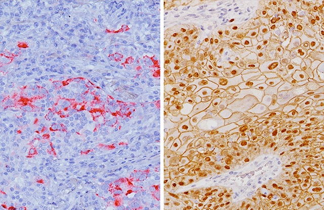
(left) and lung squamous cell carcinoma (right) stained with Desmoglein (+),
p40 (+) and Napsin A (-)
References
1. Ao, M. H., Zhang, H., Sakowski, L., Sharma, R., Illei, P. B., Gabrielson, E., Askin, F., & Li, Q. K. (2014). The utility of a novel triple marker (combination of TTF1, napsin A, and p40) in the subclassification of non-small cell lung
cancer. Human pathology, 45(5), 926–934. https://doi.org/10.1016/j.humpath.2014.01.005
2. Buxton RS, Magee AI. Structure and interactions of desmosomal and other cadherins. Semin Cell Biol. 1992 Jun; 3(3):157-67.
3. North AJ, et al. Molecular map of the desmosomal plaque. J Cell Sci. 1999 Dec; 112 (Pt 23):4325-36.
4. Savci-Heijink CD, et al. The role of desmoglein-3 in the diagnosis of squamous cell carcinoma of the lung. Am J Pathol. 2009 May; 174(5):1629-37.
5. Brown AF, et al. Tissue-preserving antibody cocktails to differentiate primary squamous cell carcinoma, adenocarcinoma, and small cell carcinoma of lung. Arch Pathol Lab Med. 2013 Sep; 137(9):1274-81.
6. Agackiran Y, et al. Desmoglein-3 and Napsin A double stain, a useful immunohistochemical marker for differentiation of lung squamous cell carcinoma and adenocarcinoma from other subtypes. Appl Immunohistochem Mol
Morphol. 2012 Jul; 20(4):350-5.
7. Bishop JA, et al. p40 (ΔNp63) is superior to p63 for the diagnosis of pulmonary squamous cell carcinoma. Mod Pathol. 2012 Mar; 25(3):405-15.
8. Tacha D, et al. An immunohistochemical analysis of a newly developed, mouse monoclonal p40 (BC28) antibody in lung, bladder, skin, breast, prostate, and head and neck cancers. Arch Pathol Lab Med. 2014 Oct;
138(10):1358-64.
9. Tacha D, et al. A six antibody panel for the classification of lung adenocarcinoma versus squamous cell carcinoma. Appl Immunohistochem Mol Morphol. 2012 May; 20 (3):201-7.


