Classification and Cocktails: Prostate Cocktail
Examining and diagnosing small prostate needle biopsies can be a difficult task. A final diagnosis of prostate carcinoma is based on
histomorphological features.1 The difficulty with examining needle biopsy stems not only from the small amount of tissue available, but also
from the fact that prostate biopsies often identify only a few malignant lesions and several histological benign “mimics”.2 These mimics
can range from seminal vesicles, Cowper’s glands, and atrophy to adenosis (atypical adenomatous hyperplasia), basal cell hyperplasia, and
even radiation atypia.1,2 Diagnosis can be complicated further when cancerous tissue is admixed with benign mimics.1 Underdiagnosis of
a small foci of malignant lesions or overdiagnosis of a cancer-mimicking benign lesions can occur and cause adverse effects for patients.
Immunohistochemistry helps in solving ambiguity and in avoiding underdiagnosis or overdiagnosis.
Biocare offers an IVD-labeled prostate cocktail (CK HMW + p63 + AMACR) that may help aid in the interpretation of troublesome prostate
lesions. The underlying concept of the prostate cocktail is to identify basal cells, found only in benign tissues, and label cancerous cells.
High molecular weight cytokeratins are expressed in a variety of normal and neoplastic epithelial tissues.3 In prostate, CK HMW [34βE12]
has been shown to be useful markers of basal cells of normal glands and prostatic intraepithelial neoplasia (PIN), a precursor lesion to
prostatic adenocarcinoma; whereas invasive prostatic adenocarcinoma typically lacks a basal cell layer.1,3,4 p63 is detected in nuclei of
the basal epithelium in normal prostate glands; however, it is not expressed in malignant tumors of the prostate.7 AMACR, also known as
α-methylacyl coenzyme A racemase, is a peroxisomal and mitochondrial enzyme that plays a role in bile acid synthesis and β-oxidation of
branched chain fatty acids.8 In immunohistochemistry, prostate glands involved in PIN have been found to express AMACR, whereas AMACR
is nearly undetectable in benign glands.9,10 Using innovative multiplex detection systems, such as Biocare’s Mach 2 Double Stain 2 polymer,
the cocktailed antibodies can be targeted and labeled simultaneously with two separate enzymes. With this polymer, the heavy weight keratins
and p63 mouse antibodies are labeled with an HRP enzyme and visualized with DAB, while the rabbit antibody AMACR is labeled with an AP
enzyme and visualized with a Fast Red chromogen.
The application of Biocare’s multiplex cocktails may be particularly beneficial in prostate cancer diagnosis. Needle biopsies supply limited
tissue available for testing. The ability for ambiguous prostate lesions to be identified and observed on a single tissue section may lead to a
successful diagnosis, as application of individual antibodies on separate slides may cause loss of atypical glands by further sectioning.1,2 It
is suggested by Chougani et al. that Biocare’s prostate cocktail can reduce the chance of overdiagnosis of benign lesions as malignant and
underdiagnosis of carcinomas as benign and reduces the chances of repeat biopsy.
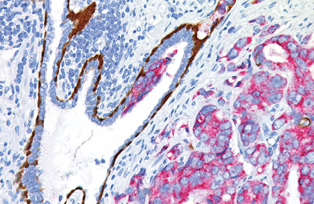
prostate cocktail.
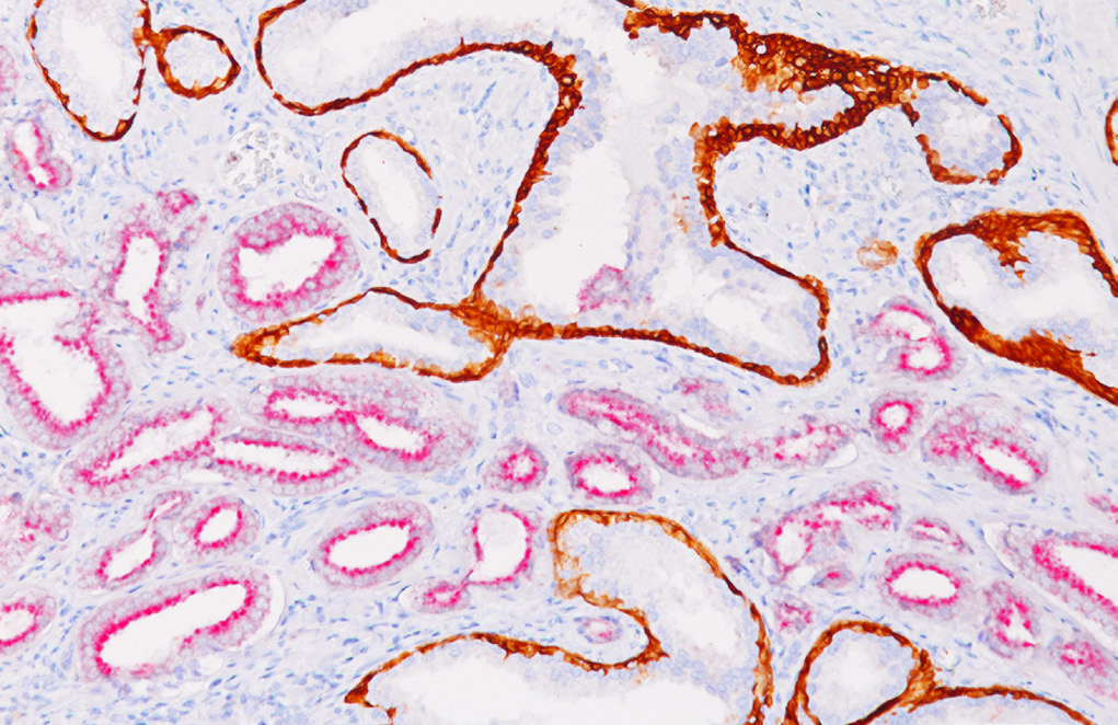
benign basal cell [brown] and prostatic adenocarcinoma [red] labeling.
Interested in accentuating your antibody menu with Biocare’s Prostate cocktail (CK HMW + p63 + AMACR)? This cocktail is available as a
standard predilute in a variety of sizes and can be used both manually and on most automated IHC instrumentation. For more information,
please call 1-800-799-9499 or visit our website: https://biocare.net/product/ck-hmw-p63-amacr-rm-2/
CK HMW + p63 + AMACR Chart
| Antibody | anti-CK HMW | anti-p63 | Anti-AMACR |
|---|---|---|---|
| Clone | 34 E12 | 4A4 | 13H4 |
| Source | Mouse Monoclonal | Mouse Monoclonal | Rabbit Monoclonal |
| Isotype | IgG1/kappa | IgG2a/kappa | IgG |
| Epitope/Antigen | CK HMW | p63 | AMACR |
| Cellular Localization | Cytoplasmic | Nuclear | Cytoplasmic |
References
1. Sujitha Chougani, Sunandalakshmi GV, Durga Kharidehal, Ravisankar V and Santhi Vissa. Utility of PIN4 cocktail antibody in the atypical foci of the Prostate. International Journal of Clinical and Diagnostic Pathology. 2020; 3(1): 396-403. https://doi.org/10.33545/pathol.2020.v3.i1f.204
2. Molinié, V., Fromont, G., Sibony, M. et al. Diagnostic utility of a p63/α-methyl-CoA-racemase (p504s) cocktail in atypical foci in the prostate. Mod Pathol 17, 1180–1190 (2004). https://doi.org/10.1038/modpathol.3800197
3. Moll R, et al. The catalog of human cytokeratins: patterns of expression in normal epithelia, tumors and cultures cells. Cell. 1982 Nov; 31(1):11-24
4. Bostwick DG, Qian J. High-grade prostatic intraepithelial neoplasia. Mod Pathol. 2004 Mar; 17(3):360-79.
5. Humphrey PA. Diagnosis of adenocarcinoma in prostate needle biopsy tissue. J Clin Pathol. 2007 Jan; 60(1):35-42.
6. Shah RB, et al. Comparison of the basal cell-specific markers, 34betaE12 and p63, in the diagnosis of prostate cancer. Am J Surg Pathol. 2002 Sep; 26(9):1161-8.
7. Signoretti S, et al. p63 is a prostate basal cell marker and is required for prostate development. Am J Pathol. 2000 Dec; 157(6):1769-75.
8. Ferdinandusse S, et al. Subcellular localization and physiological role of αmethylacyl-CoA racemase. J Lipid Res. 2000 Nov; 41(11):1890-6.
9. Zhou M, et al. Alpha-Methylacyl-CoA Racemase A Novel Tumor Marker Overexpressed in Several Human Cancers and Their Precursor Lesions. Am J Surg Pathol. 2002 Jul; 26(7):926-31.
10. Wu CL, et al. Analysis of alpha-methylacyl-CoA racemase (P504S) expression in high-grade prostatic intraepithelial neoplasia. Hum Pathol. 2004 Aug; 35(8):1008-13.


