Classification and Cocktails: Melanoma Cocktails
The skin is the largest human body organ. It is comprised of many different layers, each with its own unique function and susceptibility to
abnormalities. Melanoma is a deadly skin disease known for its aggressive clinical behavior and high tendency to metastasize.1 Most melanomas
can be accurately diagnosed by examining histological features, including asymmetry, lack of circumscription, impaired maturation, hyper
cellularity, cytological atypia, dermal mitoses, and pagetoid spread.2 The distinction of benign abnormalities from melanoma is typically
easy to establish, however, there is a subset of melanocytic tumors known for diagnostic challenges, development of late metastases, and
difficulties in treatment.1 These lesions include atypical spitzoid melanocytic proliferations, spindle cell melanomas mimicking atypical
fibroxanthomas or other fibrohistiocytic lesions, nevoid melanomas, proliferative nodules versus melanoma in large congenital nevi, melanoma
versus clear cell sarcoma, or other types of atypical nevus.2 Spitzoid proliferations are one of the hardest to diagnose.2 Research has led to the
discovery of many immunohistochemistry (IHC) markers to help delineate melanoma diagnosis.
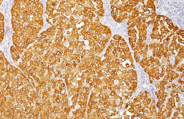
Biocare Medical offers a wide range of melanoma markers in single primary antibody and cocktailed antibody formats. Several of these
melanoma cocktails include the combination of MART-1 (Melanoma Antigen Recognized by T cells 1) and tyrosinase, as MART-1 is specific
for melanocytic lesions3-4 and tyrosinase is involved in the regulation of melanogenesis in melanocytes.1 In many melanomas, MART-1 is
co-expressed with HMB45; however, studies have shown that MART-1 is more sensitive than HMB45 when labeling metastatic melanomas.5
Additionally, tyrosinase has demonstrated to be a more sensitive marker when compared to HMB45 and MART-1 and also labels a higher
percentage of desmoplastic melanomas than HMB45.3 The combination of MART-1 and tyrosinase may also aid in identifying metastatic
melanoma in sentinel lymph nodes.6 SOX10 is another marker widely used in dermatopathology, as SOX10 has been expressed in the vast
majority of desmoplastic and spindle cell melanomas and 100% of nevi.7,8 Research suggest the combination of SOX10 with MART-1 and/or
tyrosinase labels a higher percentage of melanomas in lymph nodes and in metastatic melanoma compared to S100.9,10 Antibody cocktails
containing PRAME may be used as an ancillary tool for melanoma margin assessment. PRAME is diffusely expressed in many primary
and metastatic cutaneous melanomas, except for desmoplastic melanomas, and may aid in the distinction between nodal nevi from nodal
metastatic melanoma.11
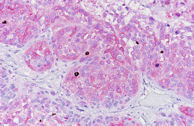
pHH3, Ki-67, and p16 are all cocktail components that may help assess melanocytic abnormalities.1 Research shows Histone H3
phosphorylation at Serine10 (pHH3) can distinguish mitosis from apoptotic nuclei, which may correlate to a type of melanoma with worse
prognosis.1,12 Adding pHH3 to a cocktail containing MART-1 and Tyrosinase may allow identification of mitotic figures with improved
specificity and time in the detection of mitotic figures in melanoma cells.1 Ki-67 is a useful marker to index proliferation.2 Researchers found
Ki-67 is highly expressed in areas of non-spitzoid melanoma, while absent in Spitz nevi.2 Combining KI-67 with Pan Melanoma (MART-1 +
Tyrosinase) serves as a tool to identify the proliferation rate of melanocytic lesions in cases in which melanocytes are sparse, there are dense
lymphocytic infiltrates, and melanocytes are admixed with fibroblasts. In general, a higher proliferative fraction is seen in melanoma than
in melanocytic nevi. p16 INK4a is a tumor suppressor protein involved in the pathogenesis of a variety of malignancies. Studies have shown
“loss of expression of the p16 in malignant melanoma was associated with tumor cell proliferation and invasive stage.”2 p16 has also proved
to be a reliable marker for the differential diagnosis between lymph node nevi and melanoma metastasis, strongly reacting in lymph node nevi
and lacking in melanoma deposits.2
The scope of melanoma malignancies might seem overwhelming. It is important to remember, however, research in IHC has provided a
wide array of diagnostic markers that may aid in the interpretation of difficult melanocytic lesions. As concluded by Shidham et al., “The
[Biocare] melanoma cocktail facilitated easy interpretation of melanoma micrometastases in sentinel lymph nodes with high interobserver
agreement. There was improvement in detection rate with the cocktail as compared to MART-1 and Melan-A individually. Furthermore,
this approach facilitates cost savings.”9 Research further suggests use of these biomarkers may aid in the identification and prognosis of
melanoma diseases, speeding up the selection of beneficial target therapy.1
Looking to expand your melanoma marker menu? Biocare’s melanoma cocktails are available as a standard predilutes in a variety of sizes
and can be used both manually and on most automated IHC instrumentation. For more information, please call 1-800-799-9499 or visit our
website: https://biocare.net/antibodies-organ/skin/.
HMB45 + Mart-1 + Tyrosinase Chart
| Antibody | anti-HMB45 | anti-MART-1 | anti-Tyrosinase |
|---|---|---|---|
| Clone | HMB45 | M2-7C10 + M2-9E3 | T311 |
| Source | Mouse Monoclonal | Mouse Monoclonal | Mouse Monoclonal |
| Isotype | IgG1/kappa | IgG2b | IgG2a |
| Epitope/Antigen | HMB45 | MART-1 | Tyrosinase |
| Cellular Localization | Cytoplasmic | Cytoplasmic | Cytoplasmic |
Mart-1 + Tyrosinase + SOX10 Chart
| Antibody | anti-MART-1 | anti-Tyrosinase | BC34 |
|---|---|---|---|
| Clone | HMB45 | M2-7C10 + M2-9E3 | T311 |
| Source | Mouse Monoclonal | Mouse Monoclonal | Mouse Monoclonal |
| Isotype | IgG2b | IgG2a | IgG1 |
| Epitope/Antigen | MART-1 | Tyrosinase | SOX10 |
| Cellular Localization | Cytoplasmic | Cytoplasmic | Nuclear |
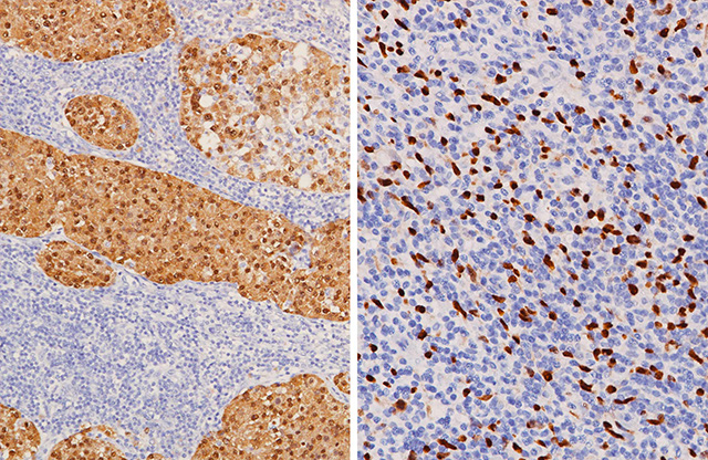
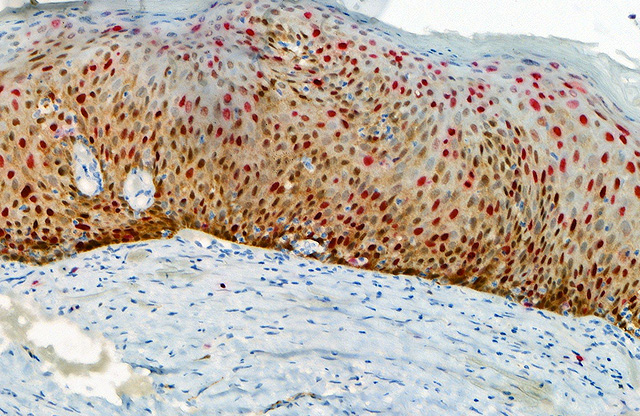
positive) and SOX10 (nuclear, positive)
(Right) Melanoma stained with MART-1 + Tyrosinase (cytoplasmic, negative)
and SOX10 (nuclear, positive).
Mart-1 + Tyrosinase + pHH3 Chart
| Antibody | anti-MART-1 | anti-Tyrosinase | BC37 |
|---|---|---|---|
| Clone | M2-7C10+M2-9E3 | T311 | BC37 |
| Source | Mouse Monoclonal | Mouse Monoclonal | Rabbit Monoclonal |
| Isotype | IgG2b | IgG2a | IgG |
| Epitope/Antigen | MART-1 | Tyrosinase | PhosphoSer10 of Histone H3 |
| Cellular Localization | Cytoplasmic | Cytoplasmic | Nuclear (mitotic figure |
Pan Melanoma + Ki-67 Chart
| Antibody | anti-MART-1 | anti-Tyrosinase | anti-Ki-67 |
|---|---|---|---|
| Clone | M2-7C10 + M2-9E3 | T311 | SP6 |
| Source | Mouse Monoclonal | Mouse Monoclonal | Rabbit Monoclonal |
| Isotype | IgG2b | IgG2a | IgG |
| Epitope/Antigen | MART-1 | Tyrosinase | Ki-67 |
| Cellular Localization | Cytoplasmic | Cytoplasmic | Nuclear |
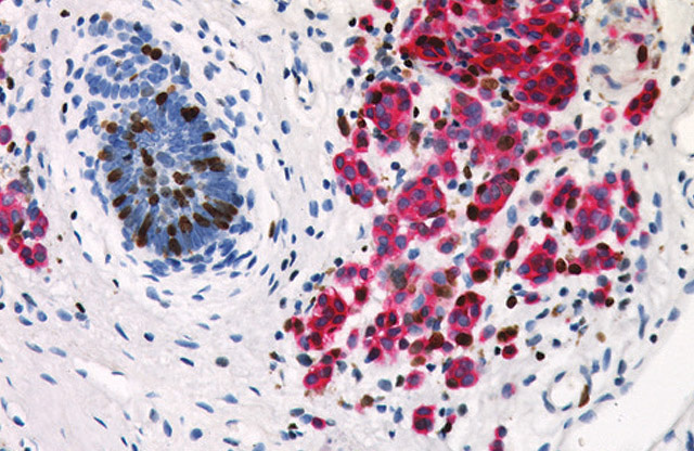
Ki-67 labeled with DAB [brown].
p16 + PRAME Chart
| Antibody | anti-p16 | anti-PRAME |
|---|---|---|
| Clone | BC42 | EPR20330 |
| Source | Mouse Monoclonal | Rabbit Monoclonal |
| Isotype | IgG1/kappa | IgG |
| Epitope/Antigen | p16 INK4a | PRAME |
| Cellular Localization | Nuclear and Cytoplasmic | Nuclear and Cell Membrane |

p16 + Ki-67 Chart
| Antibody | anti-p16 | anti-Ki-67 |
|---|---|---|
| Clone | BC42 | SP6 |
| Source | Mouse Monoclonal | Rabbit Monoclonal |
| Isotype | IgG1/kappa | IgG |
| Epitope/Antigen | p16 INK4a | Ki-67 |
| Cellular Localization | Nuclear and Cytoplasmic | Nuclear |
research suggests, an increase in p16 labeling with a decrease in Ki-67 labeling
indicates lesions may be nevus in nature.2
References
1. Tetzlaff, M. T., Torres-Cabala, C. A., Pattanaprichakul, P., Rapini, R. P., Prieto, V. G., & Curry, J. L. (2015). Emerging clinical applications of selected biomarkers in melanoma. Clinical, cosmetic and investigational dermatology, 8, 35–46. https://doi.org/10.2147/CCID.S49578
2. Botti, G., Marra, L., Anniciello, A., Scognamiglio, G., Gigantino, V., & Cantile, M. (2015). Immune-phenotypical markers for the differential diagnosis of melanocytic lesions. International journal of clinical and experimental pathology, 8(9), 9742–9751.
3. Orchard G. Evaluation of melanocytic neoplasms: application of a pan-melanoma antibody cocktail. Br J Biomed Sci. 2002;59(4):196- 202.
4. Blessing K, et al. Comparison of immunohistochemical staining of the novel antibody Melan-A with S100 protein and HMB-45 in malignant melanoma and melanoma variants. Histopathology. 1998 Feb; 32 (2):139-46.
5. Cook MG, et al. The development of optimal pathological assessment of sentinel lymph nodes for melanoma. J Pathol. 2003 Jul;200(3):314-9.
6. Miettinen M, et al.. Microphthalmia transcription factor in the immunohistochemical diagnosis of metastatic melanoma: comparison with four other melanoma markers. Am J Surg Pathol. 2001 Feb;25(2):205-11.
7. Mohamed A, et al. SOX10 Expression in malignant melanoma, carcinoma, and normal tissues. Appl Immunohistochem Mol Morphol. 2013 Dec; 21(6):506-10.
8. Mollaaghababa R, Pavan WJ. The importance of having your SOX on: role of SOX10 in the development of neural crest-derived melanocytes and glia. Oncogene. 2003 May 19; 22(20):3024-34.
9. Tacha D, et al. An Immunohistochemical Comparison Study of SOX10, Pan Melanoma Cocktail and S100 in Malignant Melanoma. American Society of Dermatopathology, 51st Annual Meeting; Poster #278, Nov 6, 2014.
10. Willis BC, et al. SOX10: a useful marker for identifying metastatic melanoma in sentinel lymph nodes. Appl Immunohistochem Mol Morphol. 2015 Feb;23(2):109-12.
11. Lezcano, C et al. PRAME Expression in Melanocytic Tumors. Am J Surg Pathol. 2018 Nov; 42(11): 1456–1465.
12. Ladstein RG, et al. Prognostic importance of the mitotic marker phosphohistone H3 in cutaneous nodular melanoma. J Invest Dermatol. 2012 Apr;132(4):1247-52.
13. LaPak KM, Burd CE. The molecular balancing act of p16(INK4a) in cancer and aging. Mol Cancer Res. 2014 Feb;12(2):167-83.
14. Shidham, V. B., Qi, D., Rao, R. N., Acker, S. M., Chang, C. C., Kampalath, B., Dawson, G., Machhi, J. K., & Komorowski, R. A. (2003). Improved immunohistochemical evaluation of micrometastases in sentinel lymph nodes of cutaneous melanoma with ‘MCW melanoma cocktail’–a mixture of monoclonal antibodies to MART-1, Melan-A, and tyrosinase. BMC cancer, 3, 15. https://doi.org/10.1186/1471-2407-3-15


