Anti-TIGIT Rabbit Monoclonal Antibody [BLR047F] – A new member of the immune checkpoint receptors family
Anti-TIGIT Rabbit Monoclonal Antibody [BLR047F]
A New Member of the Immune Checkpoint Receptors Family
Antibodies targeting immune checkpoint receptors, like cytotoxic T lymphocyte antigen-4 (CTLA-4) and cell death protein-1 (PD-1), have recently
been utilized in novel immunotherapies as they have demonstrated remarkable clinical efficiency in different tumor types, such as metastatic
melanoma, lung cancer, renal, and bladder carcinoma.1 Because of its success, blockading of other inhibitory immune checkpoint receptors is
being explored in hopes of providing further therapeutic options. T-cell immunoglobulin and immunoreceptor tyrosine-based inhibitory domain
(TIGIT), a co-inhibitory transmembrane glycoprotein receptor expressed in various T-cell subtypes and natural killer (NK) cells, is an interesting
new target for cancer immunotherapies.
“Tumor infiltrating lymphocytes (TILs) expressing TIGIT have been demonstrated in several tumor types
such as non-small cell lung cancer, colorectal carcinoma, melanoma, and acute myeloid leukaemia.1”
Recent studies have also shown a staining pattern correlation between TIGIT and PD-1, CD4, CD8, FOXP3 as well as other T-cell and NK cell
markers.1,2 Biocare Medical’s newly released TIGIT [BLR047F] specificity was evaluated based on multiplex IHC expressions in various T-lymphocytes,
NK cells, macrophages, and dendritic cells on tonsil tissue. Co-expression level between TIGIT and utilized antibodies is shown in Table 1.
Table 1
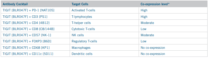
*Legend: High – >50% stained cells exhibit co-localization; Moderate – 10-49% stained cells exhibit co-localization; Low – <10% stained cells exhibit co-localization.
In this study, TIGIT [BLR047F] expressed a correct staining pattern and proved to be highly specific and sensitive to T-lymphocytes and NK
cells. High level of co-localization of TIGIT and PD-1 antibodies may suggest a co-regulatory function in immune response. TIGIT plays a crucial
role in inhibiting the tumor-directed immune response and is a valuable and competitive addition to the current array of antibodies for target
immunotherapy research.
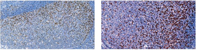
TIGIT (RM) [BLR047F] on tonsil TIGIT (red) + PD-1 (brown) on tonsil
To evaluate TIGIT in your lab, contact Biocare Medical at 800-799-9499 or visit our website www.biocare.net/product/tigit-antibody/
References: 1. Blessin N. et al. Patterns of TIGIT expression in lymphatic tissue, Inflammation and cancer. Disease Markers. Vol. 2019. 2. Kurtulus S. et al. TIGIT predominantly regulates the immune response via regulatory T cells.
J Clin Invest. 2015; 125:4053–4062.


