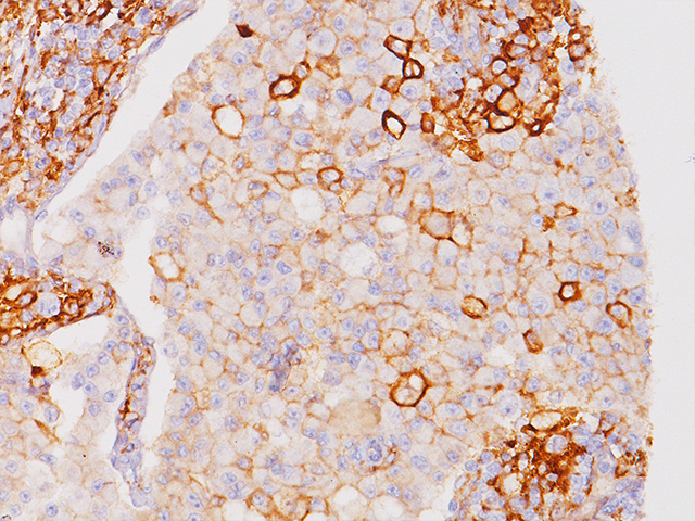
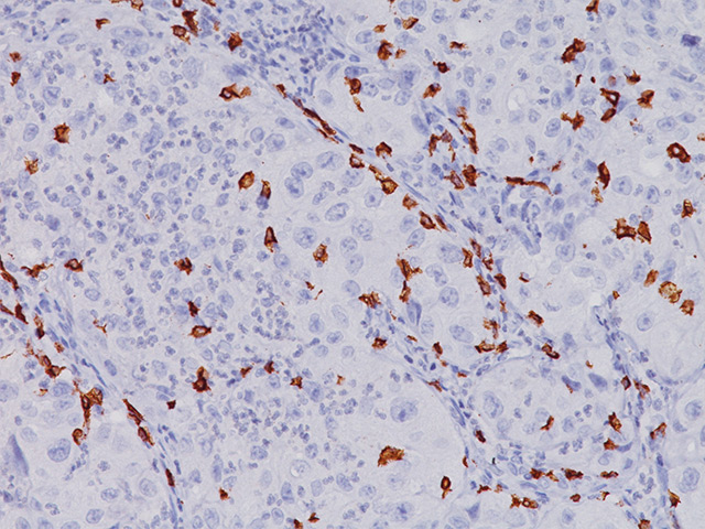
CD138/syndecan-1 is an excellent marker for identifying plasma cells, as CD138 is a transmembrane heparin proteoglycan present on the surface membrane of plasma cells that remains active in formalin-fixed paraffin-embedded bone marrow sections. Other hematopoietic cells, endothelial cells and lymphoplasmacytoid lymphomas in bone marrow are CD138 negative (1). CD138 is also expressed in fibroblasts, keratinocytes […]
Monoclonal antibody to human CD3, when used in conjunction with other antibodies, is regarded as a reliable pan T-cell antibody used in the immunophenotyping of lymphomas in paraffin sections. Most T-cell lymphomas show positivity for CD3. Notable exceptions include some of the more aggressive large T-cell lymphomas and anaplastic large cell (Ki-1, CD30) lymphomas, which […]
Programmed death 1 (PD-1) is a cell surface co-receptor in the CD28/CTLA-4 T-cell family and functions as a down regulator of the immune system through a dual mechanism of inhibition (1). PD-1 is expressed on the cell surface of activated T- and B-cells. Anti-tumor immunity may be controlled by the PD-1/PD-L1 signaling pathway. PD-L1, one […]
The prostein gene encodes a 553-amino acid type IIIa plasma membrane protein with a cleavable signal peptide and 11 transmembrane-spanning regions (1). IHC analysis has demonstrated that the Prostein antibody (also known as P501S) was expressed in the normal prostate tissue and the vast majority of malignant prostatic tissues, regardless of grade and metastatic status […]
Tumor cells can express the PD-L1 protein to protect themselves from immune system attack by binding to the PD-1 receptor on T cells, which suppresses the T cells’ ability to destroy the tumor.15 PD-L1 is frequently over expressed in placenta, and many human tumors such as melanoma, diffuse large B-cell lymphoma, and carcinomas of the […]
FOXP3 is a forkhead transcription factor family member involved in T-cell regulation, activation and differentiation. FOXP3 has been shown to be a master control gene for the development and function of CD4 /CD25 regulatory T-cells. In IHC, FOXP3 has been shown to be a specific marker for adult T-cell leukemia/lymphoma (1). In melanoma and in […]
The prostein gene encodes a 553-amino acid type IIIa plasma membrane protein with a cleavable signal peptide and 11 transmembrane-spanning regions (1). IHC analysis has demonstrated that the Prostein antibody (also known as P501S) was expressed in the vast majority of normal and malignant prostatic tissues, regardless of grade and metastatic status. Prostein expression was […]
CD4 is expressed in a T-cell subset (helper/inducer) and is found in approximately 80% of thymocytes and in 45% of peripheral blood lymphocytes. CD4 is expressed in the majority of T-cell lymphomas including mycosis fungoides, a common form of cutaneous T-cell lymphoma (1). CD8 has been shown to be an important marker in the analysis […]
The CD8 antibody reacts with the 32 kDa CD8 protein. CD8 stains cells with cytotoxic activity, including cortical thymocytes, cytotoxic/suppressor T-cells and a subset of natural killer cells. CD4 and CD8 positive and negative staining are indicative of T-cell neoplasms (1-2). CD4 and CD8 may also be used to differentiate between mycosis fungoides and cutaneous […]
CD3 is expressed throughout the T-cell differentiation process. CD3 is a highly specific and sensitive T-cell lineage marker, making it ideal for the immunophenotypic analysis of lymphohaematopoietic malignancies. Notable exceptions include some of the more aggressive large T-cell lymphomas and CD30 (Ki-1) positive anaplastic large cell lymphomas, which may not express detectable antigen.15-17 CD3 [LN10] […]


![Spleen stained with CD3 [BC33]](https://biocare.net/wp-content/uploads/3170_CD3BC33-in-Spleen-10X.jpg)