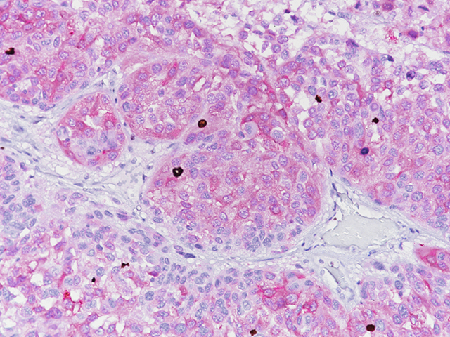
The BC23 antibody recognizes human and mouse MLH1 (80-85 kDa). The repair of mismatch DNA is essential to maintaining the integrity of genetic information over time. An alteration of microsatellite repeats is the result of slippage owing to strand misalignment during DNA replication and is referred to as microsatellite instability (MSI) (1-3). These defects in […]
MART-1 recognizes a protein of 18 kDa, identified at MART-1 (Melanoma Antigen Recognized by T cells 1). MART-1 is a useful addition to melanoma panels as it is specific for melanocytic lesions (1- 3). Studies have shown that MART-1 is more sensitive than HMB45 when labeling metastatic melanomas (3). Tyrosinase is a key enzyme involved […]
ETV6 (12p13) Orange FISH Probe is intended to hybridize to the 12p13.2 probe region on chromosome 12. The clinical interpretation of any positive or negative hybridization events should be complemented by the use of appropriate controls and other diagnostic tests where appropriate. Evaluation should be carried out within the context of the patient’s clinical history […]
Distinguishing between invasive urothelial carcinoma (UC) from other genitourinary malignancies, such as prostatic and renal carcinomas, can be difficult. Several new markers such as GATA binding protein 3 (GATA-3), S100P, and uroplakin III have been developed for this differentiation (1-7). More recently uroplakin II has been introduced (1-7). GATA-3 is a transcription factor and belongs […]
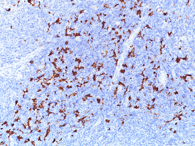
Indoleamine 2,3-dioxygenase 1 (IDO1) is a 403 amino acid cytoplasmic enzyme encoded by the INDO gene on human chromosome 8p22. IDO1 expression can be seen mainly in dendritic cells (DCs), such as immature and mature DCs, germinal center follicular DCs, interdigitating DCs, plasmacytoid DCs and in normal lymphoid organs, including lymph nodes, spleen, tonsils, Peyer’s […]
Cytotoxic lymphocytes, including NK cells and cytotoxic T lymphocytes (CTLs), play a major role in the defense against neoplastic processes and viral infections (1). Granule exocytosis is the mechanism by which cytotoxic lymphocytes may induce lysis of its target. This involves granule-associated cytotoxic proteins, including the T-cell intracellular antigen-1 (TIA-1), perforin and granzyme B (1,2). […]
CK5 and CK14 are high molecular weight cytokeratins expressed in a variety of normal and neoplastic epithelial tissues (1). In prostate tissue, mRNA for CK5 and CK14 has been detected in the basal cells of normal glands and prostatic intraepithelial neoplasia (PIN), a precursor lesion to prostatic adenocarcinoma; however, expression of CK5 or CK14 was […]
MART-1 recognizes a protein of 18 kDa, identified at MART-1 (Melanoma Antigen Recognized by T cells) (1). MART-1 is a useful addition to melanoma panels as it is apparently specific for melanocytic lesions (1,2). Studies have shown that MART-1 is more sensitive than HMB45 when labeling metastatic melanomas (3). This MART-1 cocktail does not stain […]
Detection of abnormal numbers and/or distribution of bone marrow (BM) plasma cells (PCs) on trephine biopsies can be of importance in the differential diagnosis of multiple myeloma (MM) and other PC neoplasms (1,2). CD138 expression in hematologic malignancy in bone marrow biopsy specimens is limited to nonneoplastic and neoplastic plasma cells and lymphoplasmacytic lymphoma in […]
IMP3 (insulin-like growth factor II mRNA-binding protein 3) is a 580 amino acid oncofetal RNA binding protein containing four K homology domains which is encoded by a 4350 bp mRNA transcript produced by the IGF2BP3 gene on chromosome 7p11.5 (1,2). Its relevance as a novel biomarker in many kinds of cancer has been recently published […]


![Colorectal carcinoma stained with MLH-1 [BC23]](https://biocare.net/wp-content/uploads/3214_MLH-1-BC23.jpg)